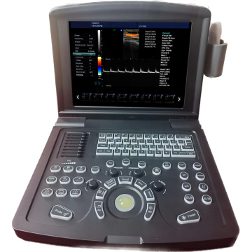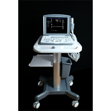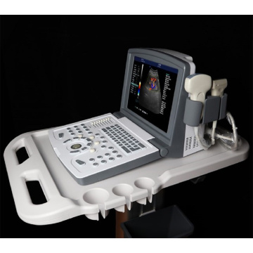


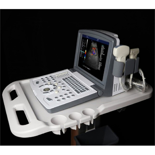
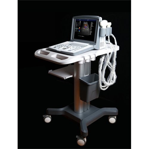
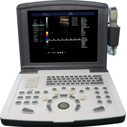
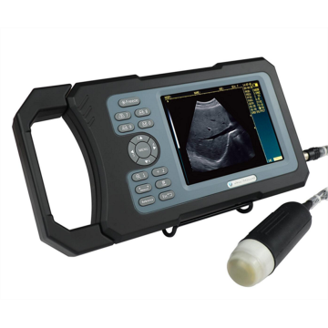
Portable Color Doppler Ultrasound Machine for Gynecology
- Min. Order:
- 5 Set/Sets
- Min. Order:
- 5 Set/Sets
- Transportation:
- Ocean, Land, Air, Express
- Port:
- Shenzhen, Ningbo, Shanghai
Your message must be between 20 to 2000 characters
Contact Now| Place of Origin: | China |
|---|---|
| Supply Ability: | 500 pieces per month |
| Payment Type: | T/T |
| Incoterm: | FOB |
| Transportation: | Ocean,Land,Air,Express |
| Port: | Shenzhen,Ningbo,Shanghai |
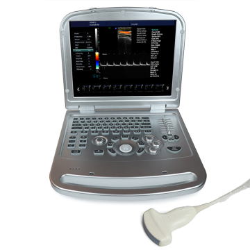
Portable Color Doppler Ultrasound Machine for Gynecology(MDK-680)
Gynecological color ultrasound mainly examines the uterus, accessories, pelvic cavity, etc.
1, uterus: color ultrasound examination can be used to determine the size of the uterus, the thickness of the endometrium and whether the echo is balanced, can also see whether there is congenital uterine malformation.
2. Accessories: Color ultrasound can detect the size of bilateral ovaries, whether there are abnormal lesions such as adjunct inflammation and ovarian cysts, and also can roughly judge the status of the fallopian tube.
3, pelvic: color ultrasound examination can also check whether there is abnormal pelvic mass, pelvic effusion, pelvic inflammation and other gynecological diseases, ultrasound accuracy is high, the operation is relatively convenient, will not cause much impact on the female body. But for abdominal color ultrasound, it is best to reserve urine in advance, so as not to affect the results of the test and thus endanger the woman's health. It is recommended to drink proper water before examination to make the bladder full to the level of the bottom of the uterus, which can make the ultrasound image clearer.
Our products:
Black and White Ultrasound Scanner
Color Doppler Ultrasound Scanner
Veterinary Ultrasound Scanner

|
No. |
Item |
Index |
|
<1> |
Depth |
≥300mm |
|
<2> |
Lateral Resolution |
≤1mm (Depth≤80mm) ≤2mm (80< Depth≤130mm) |
|
<3> |
Axial Resolution |
≤1mm (Depth≤80mm) ≤2mm (80< Depth≤130mm) |
|
<4> |
Blind Area |
≤4 mm |
|
<5> |
Geometry Position Precision |
horizontal≤5% vertical≤5% |
|
<6> |
Language |
English/Chinese |
|
<7> |
Channels |
32 |
|
<8> |
Displayer |
12” LED |
|
<9> |
External Display |
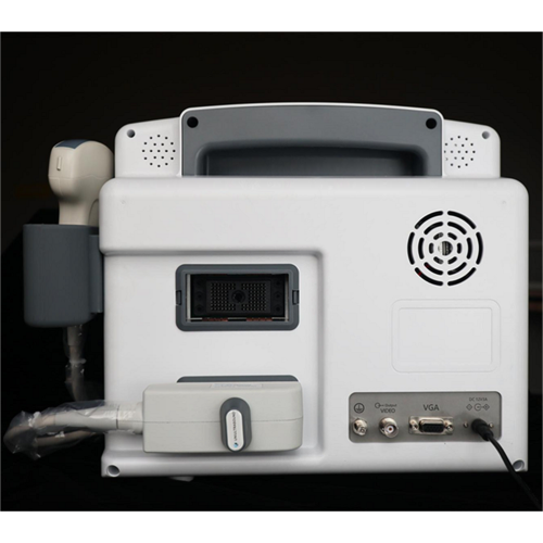
PAL, VGA, USB |
|
<10> |
Gray Scale |
256 levels |
|
<11> |
Voltage |
AC220V ±10% |
|
<12> |
Operating System |
Windows 7 |
|
<13> |
Scanning Mode |
B, B/B, 4B, B/M, M, B+C, B+D, B+C+D, PDI, CF, PW |
|
<14> |
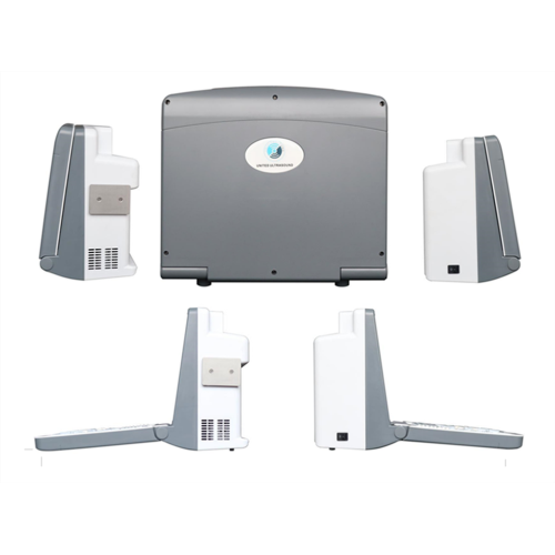
Probe |
Probe sockets: 2 Probe frequency: 2.0MHz ~ 10.0MHz, 8-step frequency conversion |
|
<15> |
Adjustment parameters of color blood flow image |
Doppler frequency, sampling frame position and size, baseline, color gain, deflection angle, wall filtering, cumulative times, etc |
|
<16> |
Signal processing |
With dynamic filtering and quadrature demodulation With total gain adjustment Gain adjustment: 8-segment TGC The total gain of Type B, Type C and Type D can be adjusted respectively B/W image gain and color blood flow gain are adjustable respectively |
|
<17> |
Doppler |
Doppler baseline adjustment level 6 Pulse repetition frequency can be adjusted separately: CFM PWD With D linear speed regulation |
|
<18> |
Digital beam forming |
Continuous dynamic focusing of digital beam forming image Full range dynamic aperture of image Dynamic tracing of the whole image Weighted Sum of Image Whole Process Receiving Delay Support half step scanning and ± 10 ° linear receiving deflection angle Multi beam parallel processing technology |
|
<19> |
Basic measurement and calculation function |
Basic measurement in mode B: distance, angle, perimeter and area, volume, stenosis rate, histogram, cross-section |
|
Basic measurement of M-mode: heart rate, time, distance and speed |
||
|
Doppler measurement: time, heart rate, speed, acceleration |
Related Keywords
-
MDK-330 Handheld Veterinary Ultrasound Scanner
MDK-380 Total Waterproof Handheld Veterinary Ultrasound Scanner
Notebook Color Doppler Ultrasonic Machine for Sheep Pig
Notebook Veterinary Black and White Ultrasound Scanner
High Quality Color doppler laptop ultrasonic machine
Related ProductsProduct Categories-
Black and White Ultrasound Scanner(21)
-
Color Doppler Ultrasound Scanner(13)
-
Veterinary Ultrasound Scanner(26)
-
Human Ultrasound Scanner(13)
-
Portable Ultrasound Scanner(15)
-
Notebook Ultrasound(15)
-
Handheld Ultrasound Scanner(13)
-
Trolley Ultrasound Scanner(12)
-
Notebook Color Doppler Ultrasound Machine(11)
-
Portable Color Doppler Ultrasound Machine(9)