

Portable Color Doppler Ultrasound Machine for Cardiac
- Min. Order:
- 5 Set/Sets
- Min. Order:
- 5 Set/Sets
- Transportation:
- Ocean, Land, Air, Express
- Port:
- Shenzhen, Ningbo, Shanghai
Your message must be between 20 to 2000 characters
Contact Now| Place of Origin: | China |
|---|---|
| Supply Ability: | 500 pieces per month |
| Payment Type: | T/T |
| Incoterm: | FOB |
| Transportation: | Ocean,Land,Air,Express |
| Port: | Shenzhen,Ningbo,Shanghai |
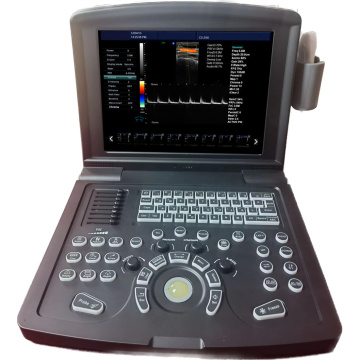
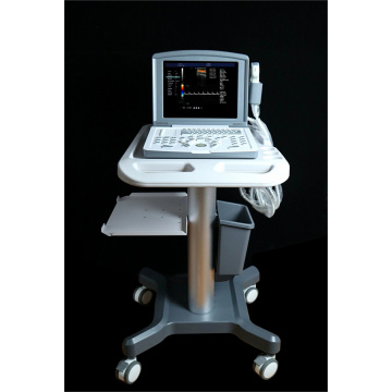
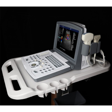
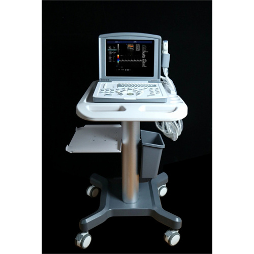
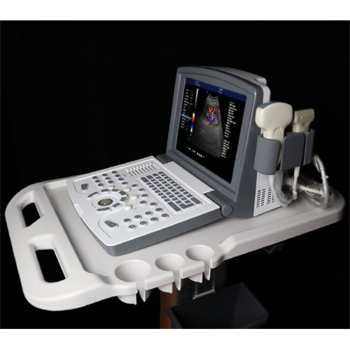
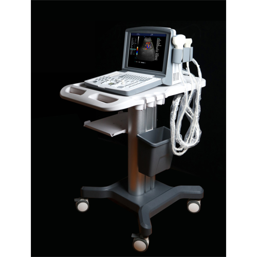
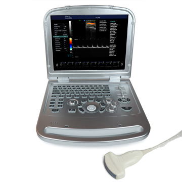
Portable Color Doppler Ultrasound Machine for Cardiac(MDK-680)
Cardiac color Doppler is a means of dynamic detection of the structure, function and blood flow of the heart using color Doppler, which only requires the use of an echocardiogram instrument. The probe is placed on the chest and viewed through different sections to obtain data on the structure and function of the heart. Including the size of the heart's chambers, such as the left atrium, the right atrium, the left ventricle, and the right ventricle. And the thickness of the left ventricular wall, both the interventricular septum and the posterior wall of the left ventricle.
In addition, the shape and structure of heart valves can be judged, including mitral, tricuspid, aortic and pulmonary valves. The systolic function of the left ventricle, that is, ejection fraction, and diastolic function of the left ventricle can also be evaluated by observing whether there are abnormalities in the shape and structure of the valve, whether there are stenosis and insufficiency, and whether there are abnormal blood flow such as regurgitation and efflux at the valve. The major arteries, such as the aorta and pulmonary artery, can also be evaluated. Therefore, cardiac color ultrasound is a very important means of cardiac examination, which can make a better evaluation of cardiac structure and function.
Our products:
Black and White Ultrasound Scanner
Color Doppler Ultrasound Scanner
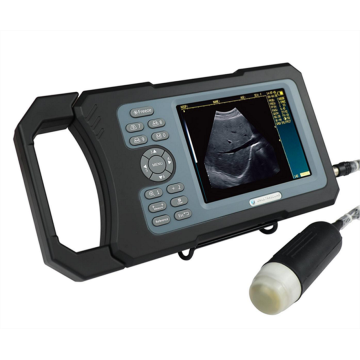
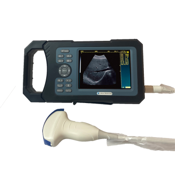
Veterinary Ultrasound Scanner

No.
Item
Index
<1>
Depth
≥300mm
<2>
Lateral Resolution
≤1mm (Depth≤80mm)
≤2mm (80< Depth≤130mm)
<3>
Axial Resolution
≤1mm (Depth≤80mm)
≤2mm (80< Depth≤130mm)
<4>
Blind Area
≤4 mm
<5>
Geometry Position Precision
horizontal≤5% vertical≤5%
<6>
Language
English/Chinese
<7>
Channels
32
<8>
Displayer
12” LED
<9>
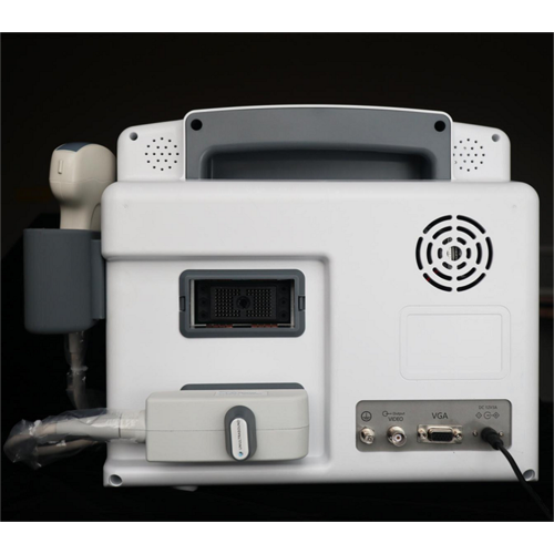
External Display
PAL, VGA, USB
<10>
Gray Scale
256 levels
<11>
Voltage
AC220V ±10%
<12>
Operating System
Windows 7
<13>
Scanning Mode
B, B/B, 4B, B/M, M, B+C, B+D, B+C+D, PDI, CF, PW
<14>
Probe
Probe sockets: 2
Probe frequency: 2.0MHz ~ 10.0MHz, 8-step frequency conversion
<15>
Adjustment parameters of color blood flow image
Doppler frequency, sampling frame position and size, baseline, color gain, deflection angle, wall filtering, cumulative times, etc
<16>
Signal processing
With dynamic filtering and quadrature demodulation
With total gain adjustment
Gain adjustment: 8-segment TGC
The total gain of Type B, Type C and Type D can be adjusted respectively
B/W image gain and color blood flow gain are adjustable respectively
<17>
Doppler
Doppler baseline adjustment level 6
Pulse repetition frequency can be adjusted separately: CFM PWD
With D linear speed regulation
<18>
Digital beam forming
Continuous dynamic focusing of digital beam forming image
Full range dynamic aperture of image
Dynamic tracing of the whole image
Weighted Sum of Image Whole Process Receiving Delay
Support half step scanning and ± 10 ° linear receiving deflection angle
Multi beam parallel processing technology
<19>
Basic measurement and calculation function
Basic measurement in mode B: distance, angle, perimeter and area, volume, stenosis rate, histogram, cross-section
Basic measurement of M-mode: heart rate, time, distance and speed
Doppler measurement: time, heart rate, speed, acceleration
Related Keywords
-
MDK-330 Handheld Veterinary Ultrasound Scanner
MDK-380 Total Waterproof Handheld Veterinary Ultrasound Scanner
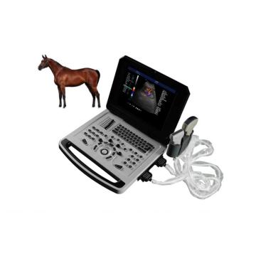
Notebook Color Doppler Ultrasonic Machine for Sheep Pig
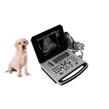
Notebook Veterinary Black and White Ultrasound Scanner
High Quality Color doppler laptop ultrasonic machine
Related ProductsProduct Categories-
Black and White Ultrasound Scanner(21)
-
Color Doppler Ultrasound Scanner(13)
-
Veterinary Ultrasound Scanner(26)
-
Human Ultrasound Scanner(13)
-
Portable Ultrasound Scanner(15)
-
Notebook Ultrasound(15)
-
Handheld Ultrasound Scanner(13)
-
Trolley Ultrasound Scanner(12)
-
Notebook Color Doppler Ultrasound Machine(11)
-
Portable Color Doppler Ultrasound Machine(9)